Appendicular Skeletal System Overview
- Identify the bones forming the pectoral girdle.
- Review and identify the bony landmarks and features of the clavicle and scapula.
- Identify the joints found at the pectoral girdle.
- Identify and review the bones of the upper limb.
- Identify the surface features and landmarks of the bones of the upper extremities.
Pectoral Girdle and Upper Extremities
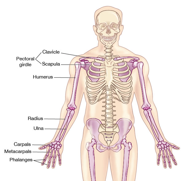
The paired pectoral (shoulder) girdles are composed of the scapulae and clavicles. These bones stabilize the shoulder and serve as anchoring points for muscles that help attach the upper extremities to the axial skeleton and provide movement at the shoulder.
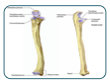
Each upper extremity includes a humerus that articulates with a scapula at its proximal end and with the bones of the forearm, the radius and ulna, at its distal end.
The radius and ulna articulate distally with the the carpals of the wrist. Distal to the carpals are the bones of the palm, the metacarpals and most distal are the phalanges, the bones found at the fingers or digits.
The muscles attached to the bones of the upper limbs provide movement at the arm, forearm, wrist, hand, and fingers.

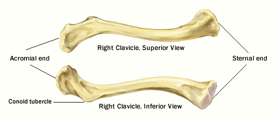
Clavicle
| Term | Definition |
|---|---|
| Acromial end | Flat, lateral end of the clavicle that articulates with the acromion of the scapula. |
| Sternal end | Medial end of the clavicle that articulates with the manubrium of the sternum; rounded to triangular in shape in cross section. |
| Conoid tubercle | Tubercle located on the inferior surface of the clavicle |
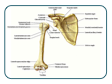
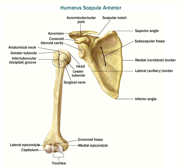
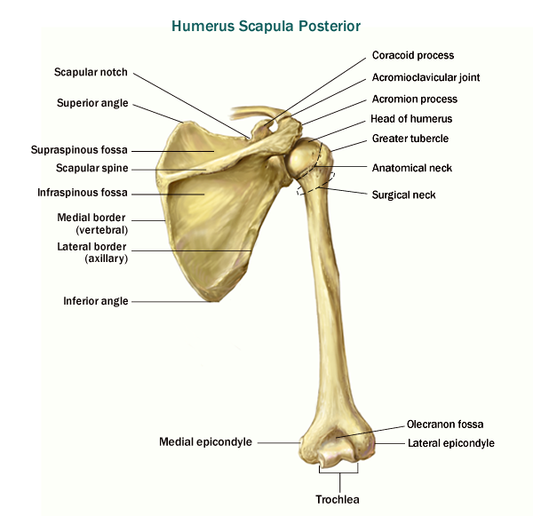
Scapula and Humerus
| Term | Definition |
|---|---|
| Scapula | Commonly referred to as the shoulder blade. |
| Acromion | Process on the lateral aspect of spine that articulates anteriorly with the acromial end of the clavicle. |
| Coracoid process | Beaklike anterior projection on the scapula; attachment site for muscles of the upper extremities. |
| Glenoid cavity (fossa) | Shallow cavity that articulates with the head of the humerus (arm bone). |
| Inferior angle | One of three angles found on the scapula; located inferiorly between the medial and lateral borders. |
| Infraglenoid tubercle | Projection located just inferior to the glenoid cavity, serving as the attachment point for the long head of the triceps brachii muscle. |
| Infraspinous fossa | Large depression located just inferior to the spine of the scapula. |
| Lateral border | The lateral portion of the scapula. |
| Medial border | The long, medial portion of the scapula, spine- sharp narrow projection on the posterior of the scapula. |
| Spine | Sharp narrow projection on the posterior of the scapula. |
| Subscapular fossa | Large depression found on the anterior surface of the scapula. |
| Superior angle | Angle between the medial and superior border of the scapula. |
| Superior border | The most superior of the scapular borders. |
| Suprascapular notch | Small notch functioning as the passageway for nerves and blood vessels located medial to the coracoid process of the scapula. |
| Supraspinous fossa | Depression located superior to the spine of the scapula. |
| Term | Definition |
|---|---|
| Anatomical neck | narrow area just distal to the head of the humerus. |
| Capitulum | Lateral condyle located distally on the humerus articulating with the head of the radius |
| Coronoid fossa | Depression on the anterior and distal aspect of the humerus. The coronoid process of the ulna fits in the coronoid fossa when the elbow is bent (flexed). |
| Deltoid tuberosity | Projection on the lateral aspect of the diaphysis of the humerus that serves as an attachment site for the deltoid muscle. |
| Greater tubercle | Large, lateral projection on the epiphysis of the humerus. head- rounded, proximal end of the humerus that articulates at the glenoid cavity of the scapula. |
| Intertubercular groove/sulcus | Groove between the greater and lesser tubercle housing the tendon for the biceps muscle. |
| Lateral epicondyle | Raised ridge proximal to the lateral condyle (capitulum). |
| Lesser tubercle | Medial projection on the epiphysis of the humerus. |
| Medial epicondyle | Raised ridge proximal to the medial condyle (trochlea). |
| Olecranon Fossa | Depression on the posterior, distal epiphysis of the humerus. radial fossa- depression on the anterior, distal aspect of the humerus, just lateral to the coronoid fossa. |
| Surgical neck | Narrowed region at the proximal end of the diaphysis. A common site for fracture. |
| Trochlea | The medial condyle of the humerus, articulating with the trochlear notch of the ulna. |
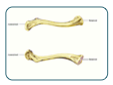
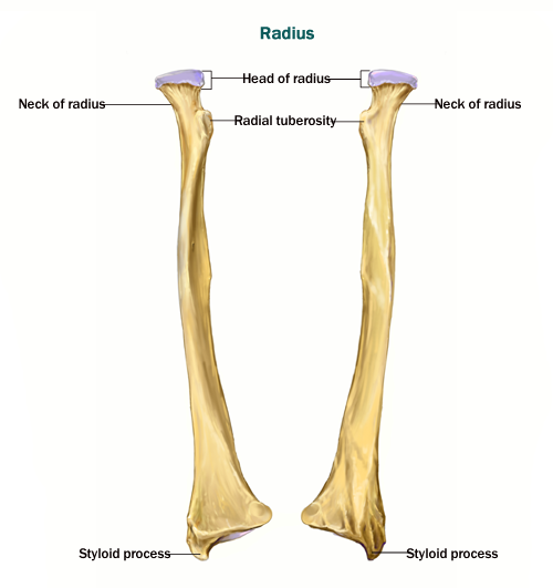
Radius (Where is Ulnar notch ?)
| Term | Definition |
|---|---|
| Radius | Lateral forearm bone (in the anatomical position) |
| Head | Proximal end of the radius articulating with the capitulum of the humerus. |
| Neck | Narrow area just distal to the head of the radius. |
| Radial tuberosity | Medial projection on the proximal radius serving as an attachment point for the biceps brachii muscle. |
| Styloid process of radius | Small projection on the distal radius. |
| Ulnar notch | Depression located on the medial aspect of the distal radius that articulates with the distal end of the ulna. |
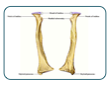
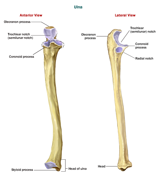
Ulna Where is Ulnar tuberosity - projection on the anterior surface of the proximal ulna that is the site of attachment for the brachialis muscle.
| Term | Definition |
|---|---|
| Ulna | Medial forearm bone (in the anatomical position) |
| Coronoid process | Small projection located on the anterior aspect of the proximal ulna, anterior to the trochlear notch, articulating with the coronoid fossa of the humerus when the elbow is bent (flexed). |
| Olecranon process | Large, posterior projection on the proximal end of the ulna serving as the attachment point for the triceps brachii muscle. |
| Radial notch | Depression on the lateral aspect of the proximal ulna where it articulates with the radius. |
| Styloid process | Small projection on the distal ulna. |
| Trochlear notch | Depression on the proximal ulna that articulates with the trochlea of the humerus. |
| Ulnar tuberosity | Projection on the anterior surface of the proximal ulna that is the site of attachment for the brachialis muscle. |
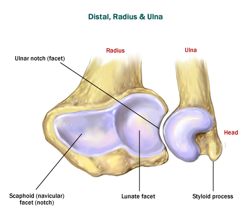
Distal End of Radius and Ulna
| Term | Definition |
|---|---|
| Radius - Lateral forearm bone (in the anatomical position) | |
| 1. Lunate facet | Surface on the distal radius that articulates with the lunate bone. |
| 2. Scaphoid (navicular) facet (notch) | Surface on the distal radius that articulates with the scaphoid bone. |
| 3. Ulnar notch | Depression located on the medial aspect of the distal radius that articulates with the distal end of the ulna. |
| Ulna - Medial forearm bone (in the anatomical position) | |
| 1. Head | Rounded eminence on the lateral surface of the distal ulna. A portion of it articulates with the radius. |
| 2. Styloid process | Small projection on the distal ulna. |
Carpals, Metacarpals and Phalanges
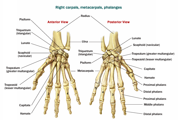
| Term | Definition |
|---|---|
| Carpals | 8 bones making up the wrist |
| Proximal row: (lateral to medial) | |
| 1. Scaphoid | S shaped carpal bone articulating with the radius |
| 2. Lunate | “Moon” shaped carpal bone that articulates with the radius. |
| 3. Triquetrum | Triangular shaped carpal bone articulating with the ulna. |
| 4. Pisiform | Small “pea” sized carpal bone located most medially. |
| Distal row: (lateral to medial) | |
| 1. Trapezium | Table-shaped bone located at the base of the first metacarpal. |
| 2. Trapezoid | Table-shaped bone located at the base of the second metacarpal. |
| 3. Capitate | Largest of the carpals, head-like in shape and located in the center of the wrist at the base of the third metacarpal. |
| 4. Hamate | Has a hook-like appearance, located at the base of the fifth metacarpal. |
| Metacarpals - bones at the palm; five in total each with the following structures: | |
| 1. Base | Proximal epiphysis |
| 2. Body | Diaphysis |
| 3. Head | Distal epiphysis |
| Phalanges - bones at the digits; I (pollex) – thumb; composed of a proximal and distal phalanx | |
| 1. Proximal | Phalanx closest to the metacarpal |
| 2. Distal | Phalanx that is triangular in shape and most distal |
| II-V- digits II-V; each composed of a proximal, middle and distal phalanx | |
| 1. Proximal | Phalanx closest to the metacarpal |
| 2. Middle | Phalanx between the proximal and distal phalanxes. |
| 3. Distal | Phalanx that is triangular in shape and most distal |